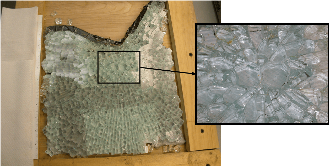
A tempered glass pane was reported to have shattered unexpectedly after approximately 30 years of service. The portion of the pane provided for analysis, shown in Figure 1, included the origin of the failure which was identifiable by the crack pattern radiating from two rather larger polygonal glass pieces forming a shape frequently referred to as a “butterfly.” This pattern is consistent with a stress concentration at the origin point.
Spontaneous failure of tempered glass, with no obvious outside cause such as mechanical impact, is typically the result of an inclusion in the glass body. Such inclusions are often nickel sulfides with or without additional contaminants present. These inclusions form due to impurities present at the time the glass is manufactured. There are a number of stable nickel -sulfide forms, the simplest of which is NiS. The NiS crystal can exist in two phases: a lower volume alpha (α) phase that is stable at higher temperatures (above 379°C) and a higher volume beta (β) phase that is stable at lower temperatures. Tempered glass is manufactured by heating annealed glass well into the α-NiS temperature range, followed by rapid quenching. The process produces a glass product with its surface under compressive stress and its interior under tensile stress. When tempered glass breaks, it is this stress arrangement that causes the characteristic pattern of many relatively small and rounded pieces, as opposed to fewer large and jagged pieces. However, any NiS impurities that may be present in the glass body can be locked in the α-NiS structure by this process. A trapped α-NiS inclusion is metastable at temperatures below 379°C and can slowly transition over time from the lower volume phase to the higher volume β -NiS structure. The volume increase may be as great as 4%, building localized stresses in the glass body that eventually lead to sudden catastrophic failure. The speed of the phase transition can be extremely slow and is affected by thermal factors such as heating by sunlight or the lack thereof.
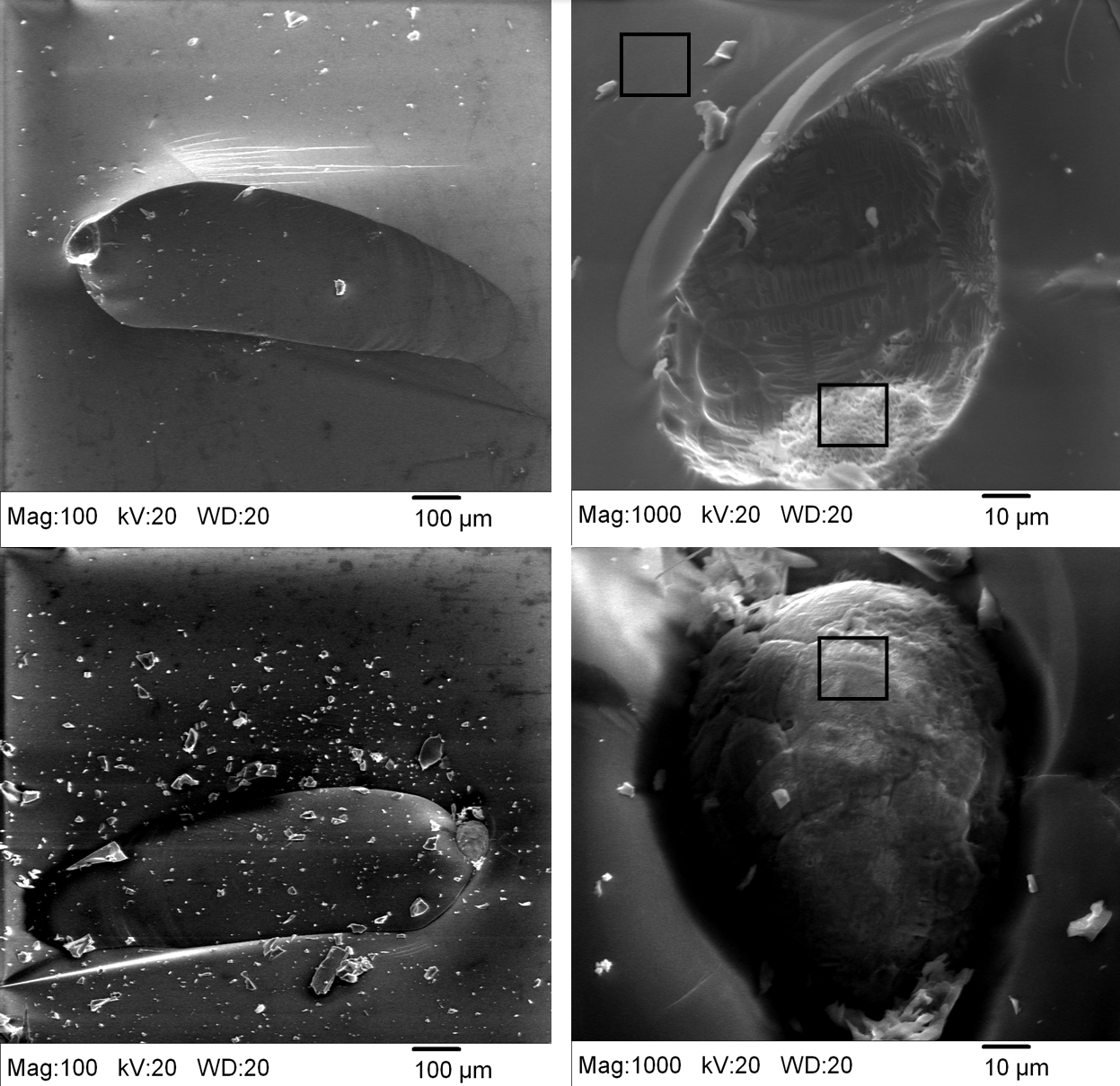
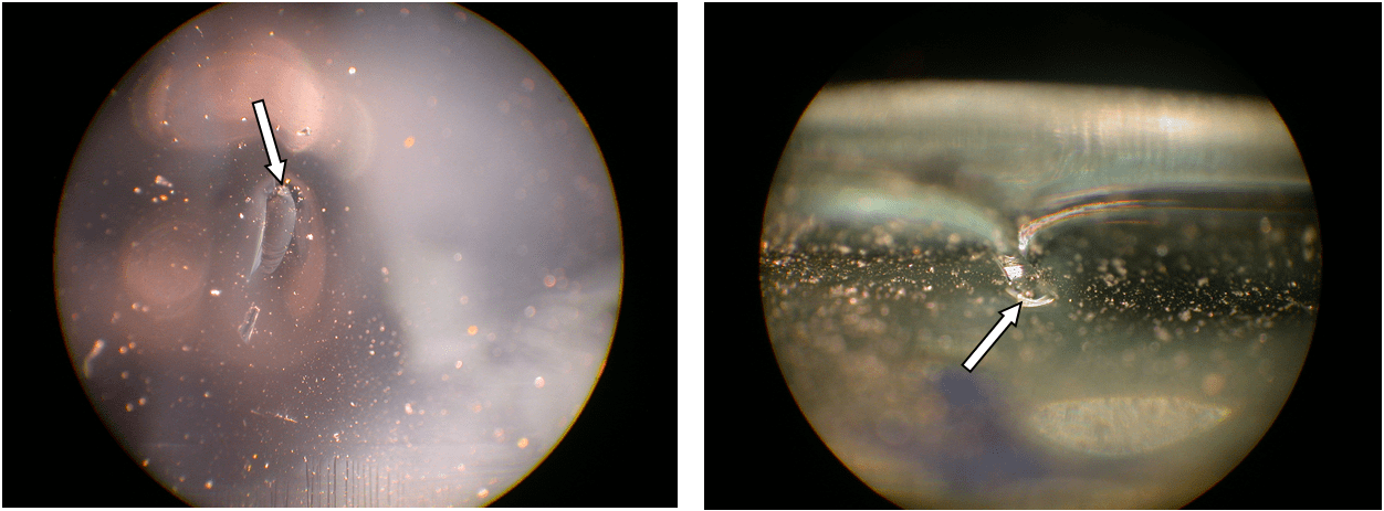
For the fracture under investigation here, the butterfly pieces were extracted from the specimen and the mating cross section surfaces were initially inspected with optical microscopy. Microscopic chip features associated with the origin of fracture were noted roughly halfway through the thickness of the pane, confirming an inclusion. Example images are shown in Figure 2.
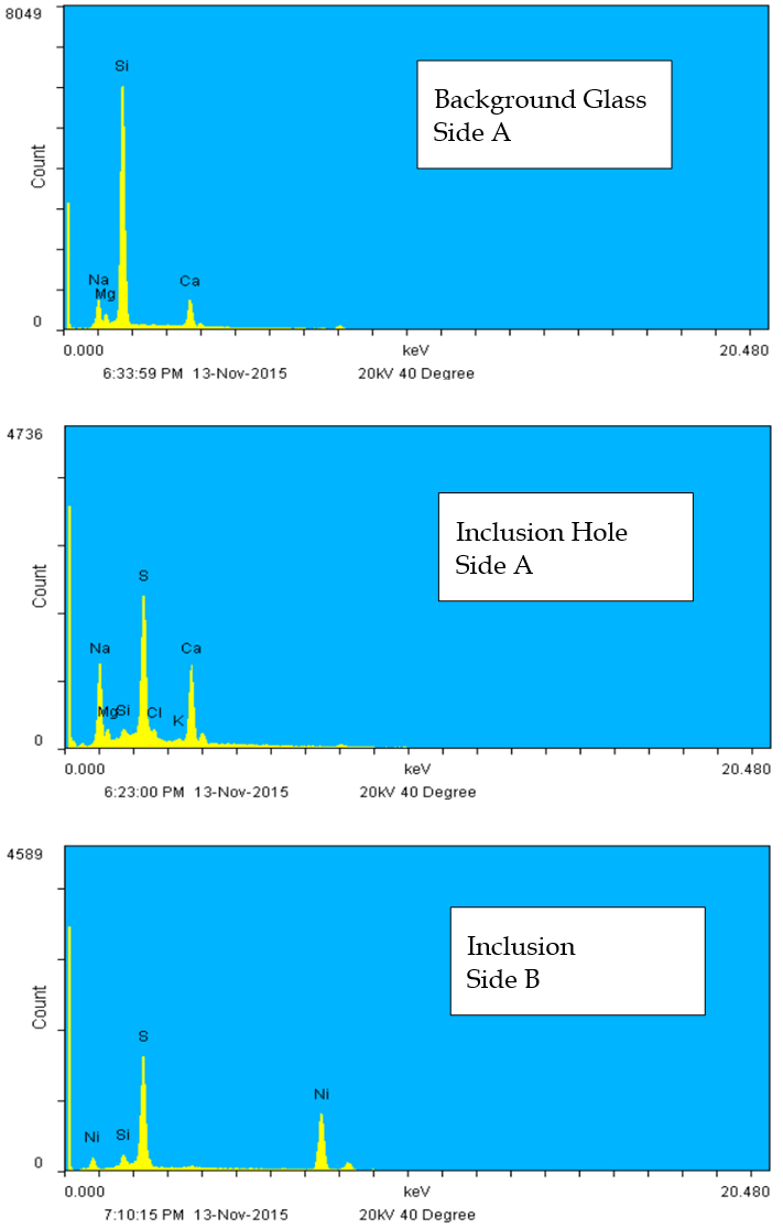
The specimens were next examined by scanning electron microscopy/energy dispersive x-ray spectroscopy (SEM/EDX). The SEM images of Figure 3 show the initial crack zone and the origin. The elliptical ball-like particle first noted by optical microscopy on “Side B” is the NiS inclusion, as revealed by EDX. The EDX spectra of Figure 4 show the elemental compositions of the background glass, the impurities at the inclusion/glass boundary (inclusion hole), and the inclusion itself. Note that only the inclusion itself contains Ni as well as S, and no obvious impurity elements are present in detectable concentrations in the glass body.
By locating the nickel sulfide inclusion at the fracture origin this failure was proven to be the result of impurities present in the glass pane at the time of manufacture.